

![]()
STANDARDIZED MEASURE OF VISUAL ACUITY
F.RICCI, C.CEDRONE and L.CERULLI
Research developed within the targeted project FATMA (Prevention and control of risk factors), sub-project Community Medicine of the CNR (Italian National Research Council), 1990-1995.
Grant n.9100119.pf41.
![]()
Summary: the Authors reviewed the main aspects in visual acuity evaluation such us the characteristics of test devices, notation employed in recording the visual acuity level and the procedures for VA assessment, both in distance and in near test. Furthermore new ten-letter charts, that tend to follow more strictly the standardization guideline suggested by NAS-NRC, are described.
Key Words: Visual acuity, optotype charts, luminance, contrast, forced choice.
![]()
The measurement of visual acuity (VA) is an essential part of the ophthalmologic examination and represents the most common and useful test for the visual function assessment. Although we usually think about the VA as a measure of the resolving power of the foveal area, assessment of abnormal visual function may be related to different conditions such as refractive errors, media opacities, retinal and optic pathway diseases.
The specific need for standards is related to the type of use to which the test is designed. The common aim is to compare repeated measurements made by more than one examiner also in different place [1].
Thus, in clinical practice the primary need is to compare the results over time to evaluate the occurrence or the evolution of pathology; in clinical trials or research projects, the need is to perform a reliable and reproducible measure; in qualifying tests (physical standards) the need is to employ univocal judgement criteria.
A recent work [2] summarizes the recommended procedures for the standardized measure of VA evolved as part of 20 years of clinical research.
The aim of the present work is to provide not only a guideline, but also a revue of the main arguments related to the standardized assessment of the VA.. Furthermore we propose new standard VA test charts designed following the basic recommendations of NAS-NRC [1], whose validation and comparison with the current standard charts are in progress.
Standardization of VA measurement is a global procedure that involves different aspects such as the characteristics of the test devices, the notation employed in recording the acuity level and the strategy for absolute threshold assessment. In the present work our attention is restricted to the "recognition acuity" (Minimum legible acuity).
![]()
Characteristics of the test devices
"Distance Acuity Test"
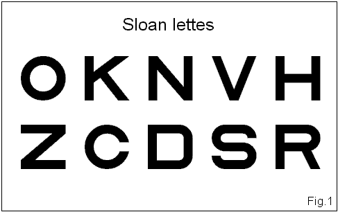
Standard optotypes: The Landolt broken rings are widely accepted as the reference standard [3]. The use of other optotypes requires a demonstrable equivalence to Landolt rings and an equal recognition difficulty. Both Sloan [4] and British Standard Institution [5] optotype sets show these features [6,7,8] (Fig.1)
Optotypes scaling:
optotypes size has to be reduced in a constant way in order to obtain an equal variation all over the scale extension [9]. A logarithmic progression in steps of 0.1 logUnits corresponds to a geometric progression in which each row contains optotypes whose size is 1,26 times smaller than the preceding row. VA threshold is obviously affected by the rate of scaling employed.Scale extension: a useful scale extension spans at least from 10’ to 0.8’ minutes of arc of MAR of stroke, at the full test distance [2,10].
Optotypes spacing: optotypes have to be quite far apart to avoid the "crowding effect" [11,12]. If this distance is at least 5 times their visual angle, the acuity may be assumed equal to that measured with isolated characters ("Interaction free acuity"). Because the strength of the crowding effect varies as a function of the distance between the optotypes, VA measure performed by means of non-homogeneous charts may lead to very different results [8].
Number of optotypes: the same number of optotypes at each size level is required; the recommended number is ten optotypes per row. Eight letters per row is the minimum accepted. It is helpful to divide the ten optotypes in two rows [1].
Background luminance: in the normally sighted, VA increases as function of the background luminance from mesopic to high photopic luminance until glaring brightness is obtained, at which VA begins to decrease [13]. VA increase spans from 0.025 cd/m2 to 60 cd/m2. Above 80 cd/m2 the increase is very slight and above 500 cd/m2 it is quite negligible [14]. This effect is related to the improvement of contrast sensitivity at high level of luminance [15].
Ambient luminance: maximal VA is achieved in photopic conditions. It has been demonstrated that keeping constant the contrast and the luminance of the test target, VA varies with the retinal adaptation conditions. In photopic adaptation the VA increases as target luminance increase. In scotopic or mesopic adaptation peak levels of VA are relatively smaller [16]. It is favourable to avoid VA measurement in a dark room but ambient luminance should not exceed one half of the chart background luminance [1].
Light wavelength: changes in wavelength of the background light source lead to image defocusing that in dynamic conditions produces a variation in amount of accommodation. In cases of achromatic light such as daylight (CIE Source 6500) incandescent lights (CIE Source A) and fluorescent tubes, it is possible to assess the wavelength for which the eye must be focused to obtain minimum blur, as well as the relative amount of defocusing for different sources [1]. The spectral centroid of a source may be assumed to correspond to the wavelength for which the eye must be focused [17]. This is true also for fluorescent sources in spite of continuous spectrum of emission [18]. Assuming that the human eye is in focus at wavelength of sodium emission (589 nm), to get a defocusing of 0.25 diopters it needs a wavelength shift of 45 nm. Light sources whose spectral centroid falls in this range (589-544 nm) are useful for background illumination of standardized chart [1].
Optotypes contrast: the capability to identify an optotype is not only related to its angular width but to the test target contrast too. VA (resolution or recognition) is reduced as the contrast between the target test and background decreases. When the contrast is reduced below a critical value (threshold) the capability to resolve the target fails [19,20,21]. The maximal VA is achieved with charts whose contrast is above 80%; the measure of the contrast of VA charts is then seldom necessary, because the charts' contrast is widely in the desired range.
Test distance:
changes in test distance in a dynamic condition alter the focus and then the related amount of accommodation. VA measurement performed at different distances may be affected by this factor. Standard chart testing distance should to be 4 meters and many arguments support this choice:2. "Near Acuity Test":
To provide measures of near acuity comparable to those obtained at four meters, the near chart should meet the same specifications outlined in the distance charts [1]. There are evidences that if the near tests corresponds in all respects to the standard employed for distance test, in a large majority of patients the threshold visual angle expressed as logMAR will agree within 0.1 logUnits. Difference in distance and near tests occurs in patients with cataract where the distance acuity is generally lower than the near one. Retinal and optic pathway diseases usually affect distance and near acuity at the same level.
![]()
Characteristics and limits of the current distance charts.
1. Snellen chart:
is one of the most widely used charts for VA testing. In spite of his wide diffusion the chart organisation shows several limits for standard measurement [7][24][25]:- different number of optotypes per row; in this condition as one reads smaller rows the task difficulty increases.
- irregular progression in letter size; the scale of the measure is not the same all along the extension of the chart so that the gain or the lost of one line has not the same value in different part of the chart.
- differences in optotypes recognition difficulty; the chart shows letters relatively easy such as A and L, and others more difficult such as B, E and F
- difference in background luminance related to different chart manufacturers.
2. ETDRS chart: it represents the major effort to introduce a standardized chart based on the recommendation of NAS-NRC. The chart, introduced by Ferrys [7] modifying the Bailey & Lovie chart [26], shows a regular progression of the type size and spacing, following a logarithmic scale in steps of 0.1 logUnits, presents five Sloan optotype per row [3]. The use of five letters per line requested to choose the combination of Sloan optotype in order to obtain the same mean difficulty for each line
(Tab1). The Authors pointed out that the use of 5 letters per row is the only deviation from the NAS-NRC Committee recommendation. The reduced number of optotypes increase the probability of false recognition due to guessing driven by the forced choice method [ 27].The background luminance is about 150 Cd/ m2 and then exceed the limit of 85
+ 5 cd/m2, and there is not any possibility of regulation. However this feature does not affect the final results of the measure, and that other Authorities suggest a standard background luminance ranging from 120 to 300 cd/m2 [5,28].
Characteristics and limits of the current near charts.
The first problem in establishing near test standards is if it is better to use a text-reading task rather than optotypes recognition. Text reading is qualitatively different from the individual optotype recognition, with weak or no correlation between the two tests. Consequently, text based testing will find a more correct employ in the evaluation of visual impairment rather than in VA measure [4,29,30,31].
A lot of optotypes based system have been proposed most of which have many flows. In fact both "Jaeger system" and "point system" can vary across different manufacturers of test card, and then, in spite of their diffusion, they are not useful in standardized assessment [32,33].
![]()
Is there room for new charts for visual acuity measurement?
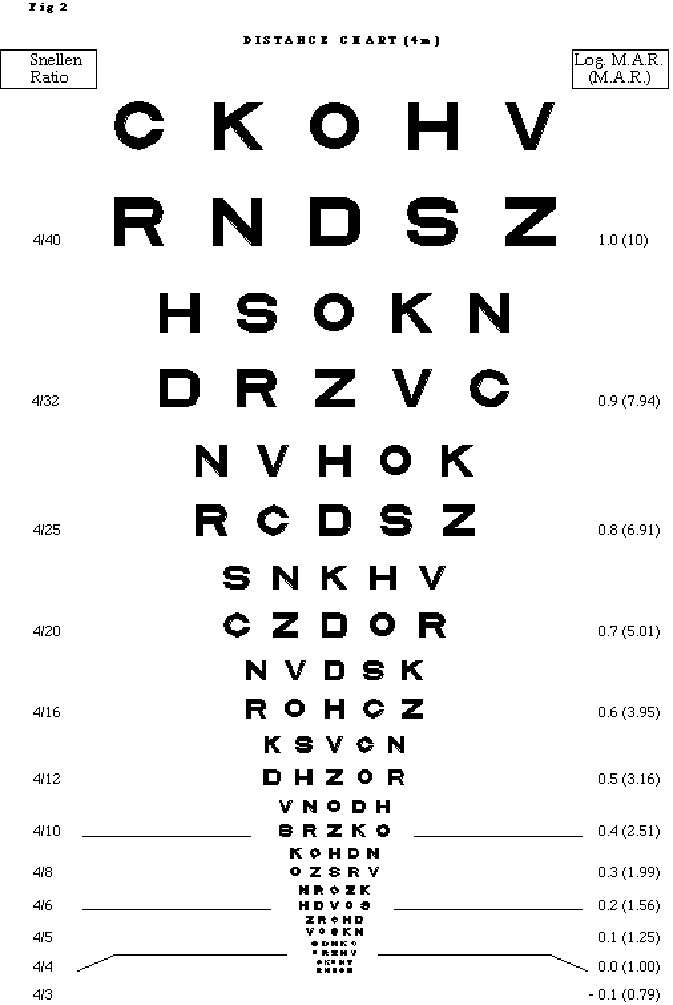
We designed new visual charts that tend to follow more strictly the standardization guideline, with aim to improve the ETDRS charts [34] (Fig.2) (Tab2).
The characteristics of the distance test chart are summarized:
Near test chart: it is a 1:10 scaled down chart. The test distance is then of 40 cm. The scale extension spans from 1.4 logMAR (4/100) to -0.1 logMAR (4/3) at full distance
(Fig.3) (Tab.3). The main differences in respect to ETDRS charts are:![]()
Notation employed in recording the VA level
Distance charts
VA = m/M
In Europe there is a strong practice to convert the Snellen ratio to the decimal system (decimal acuity) [35]. In our Country further approximation is performed transforming the decimal acuity in decimal ratio, where 10/10 means both 6/6 and 5/5 and 4/4 and 3/3 acuity, and 1/10 means 6/60 or 5/50 etc. Both decimal acuity and decimal ratio allows direct comparison of VA results performed both with 3, 4, 5 and 6 meters charts, but they represent an improper use of Snellen ratio [36]. Decimal acuity may be confused both with the Snellen Sterling "percentage of visual efficiency" which has a very different meaning and with the MAR values [15,37].
- States VA in absolute terms
- Does not involve any assumption of normal reference values
- Can be used with charts of any letter size progression
- Allows direct comparison of values obtained with different distance charts
- Allows direct comparison between distance and near acuity level
- Allows easy conversion from and to Snellen notation.
- Is expressed as measure unit of international employ, and is then useful to definition of an international standard.
- Easy progression in steps of 0.1, from +1 to - 0.1 (-0.2)
- Capability to express VA as an interpolated value [6][39].
Near charts
VA = c/C
![]()
STANDARD PROCEDURE FOR VA ASSESSMENT.
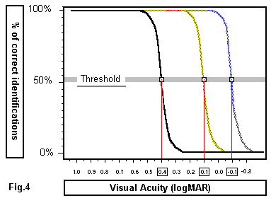
Forced choice: the general purpose of psychophysical method is the measurement of the threshold. This term refers to a boundary stimulus that results in a change from "sensation" to "no sensation". When we evaluate VA level we detect the minimum width of the stroke expressed in minutes of arc that allows the correct identification of the test ("absolute threshold") [41]. The threshold is defined statistically as the boundary stimulus recognized more than 50% of the number of its presentations (Fig.4).
Subjects with the same recognition acuity may perform different results to the VA testing in order to the aptitude to answer when tested with threshold stimuli. Forced choice procedure can limit differences related to the individual response bias or criterion [42]. This method requires that the percentage of the identifications have to be corrected for guessing. The following relation, called Abbot formula, allows calculating the number of identifications corrected for guessing [43,44] :
CI = N (n-1) - Mn / N (n-1)
In the formula CI is the percentage of correct identifications, n is the number of optotypes per row, N is the number of presentations and M is the number of mistakes.
Table 4 shows the percentages of correct identifications calculated in different test conditions.
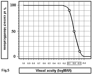
Stating exam end point. There is a close relationship between VA threshold and criteria applied to evaluate subject performance in reading an eye chart. The plot of a frequency seeing curve indicates that VA threshold varies according to the percentage of correct responses assumed as end point (Fig.5).
The ophthalmic literature shows different criteria for measuring visual. Most of the Authors commonly used as end point the correct identification of 50%+1 of the character of a defined line on Snellen chart. This criterion was employed in the Framingham Eye Study [45]. Ferris suggested the identification of 4 out of the 5 characters per line in charts arranged in logarithmic scale [7]. Wong e Kaye suggested for acuity screening the correct identification of 100% of characters in chart arranged with 2 letters per row, especially designed for quick test [46]. The NAS-NRC recommended standard is the correct identification of 50% + 2 of the characters for charts with 10 optotypes per row. In this way the probability of guessing 7 correct identification out of 10 letters is less than 1% and then the measure of VA is highly significant [1].
LogMAR notation allows the recording of VA level in terms of interpolated values. We can assign to each optotype a score that is equal to the value of the logarithmic progression (0.1) divided by the number of the optotypes per angular width. In a ten letter chart this value is then 0.01(0.1/10) per recognised optotype. If a patient can read 7 out of ten letters of the row corresponding to 0.0 LogMAR and two letters of the next -0.1 line, we can interpolate a VA score subtracting 0.01 per recognised letter in the next row. In this example visual acuity is "VA = 0.0 - 2 x 0.01= -0.02", that represents a score interpolated between 0.0 and -0.1 logMAR.
Acuity score may be useful both in detecting subtle change in VA and in performing accurate statistic tests [7].
Standard methods for measuring low VA.
The reduction of VA below 4/40 requires that VA testing is performed at a distance closer than standard one.With a 4 meters logarithmic chart we can follow two procedures related to the format of the notation employed.
1) If we use the MAR notation or the Snellen ratio, reducing to a half the test distance we double the MAR resolution or double the denominator of Snellen Ratio.
Es: 4/40 at 4 meter = 4/80 at 2 meters = 4/160 to one meter etc.
2) If we employ the logMAR format we can reduce test distance in progression of 0.1 logUnits (4 - 3.2 - 2.5 - 2.0 - 1.3 and 1). Each step of reduction corresponds to the variation of +0.1 logUnit on the chart scale. Identification of the first line corresponds to VA of logMAR +1.0 at 4 meters, +1.1 logMAR at 3.2 meters, +1.2 logMAR at 2.5 meters etc.
(Tab.5) (Fig 6).Different charts for refraction and VA measurement.
In order to avoid memorization of the sequence of the optotypes it is strongly recommended to employ at least 3 different charts to perform the distance acuity test. The first chart must be used only for refraction, the second for VA assessment in the right eye and the third for the left eye. Likewise near acuity must be performed with different charts for each eye, using a lighting device that allows performing the measure at the same background luminance employed in distance test.
![]()
Discussion.
Snellen chart, in spite of its diffusion, presents too many flaws to represent the reference one. Charts with a regular progression of the type size and spacing, with the same number of letters per row and equal recognition difficulty (Landolt C or Sloan letters) are more useful for the standardization requirements.
VA test is currently employed for different purpose ranging from the assessment of refraction to the evaluation of visual function. Wong e Kaye [45] suggested that different charts may be useful for different purposes and each chart should balance sensitivity specificity and requested time in relation to specific needs.
High sensitive test charts produce a low percentage of false negatives, while high specific tests produce few false positive responses. Sensitivity of the chart is related both to the number of the letters per row and to the requested number of correct identification for VA threshold identification. Specificity of the test may be reduced by difficult and time-consuming exams.
A two-letters chart joint to a suprathreshold endpoint may be useful in VA screening while large epidemiological population studies may benefit from more balanced test such as ETDRS five-letters charts (7).
Nevertheless in other studies, such as refractive surgery trials, cataract medical treatment trials etc. it may be relevant to perform the measure of VA threshold or score in order to evaluate small differences overtime. In these cases the use of the described ten-letter chart may allow to perform more sensitive and specific tests.
![]()
References
![]()